VUV

Beamline VUV-Photoemission

Paolo Moras  - paolo.moras@ism.cnr.it

Laboratory: Elettra

 
The VUV-Photoemission beamline at the synchrotron Elettra (Trieste) is dedicated to the study of the electronic and magnetic structure of solid systems by photoemission spectroscopy. The valence band, Fermi surface and core levels of these systems, which define their chemical, electronic, optical and transport properties, can be analyzed in detail owing to the unique combination of high brilliance and tunability of the synchrotron light and the high energy and angle resolution provided by the Scienta R4000 electron spectrometer. Samples can be studied over a wide range of temperatures and using different scanning geometries, thanks to the 5-degree of freedom cryo-manipulator. The data acquisition setup is automatized and employs user-friendly software. The preparation and structural characterization of the samples can be performed in-situ under ultra-high vacuum conditions by sputtering, annealing, cooling, epitaxial deposition of various materials, exposure to gases, and low-energy electron diffraction.

Further information at: http://www.elettra.eu/elettra-beamlines/vuv.html


TECHNICAL SPECIFICATIONS
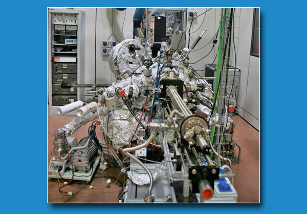
Beamline

  • Photon energy: 20-750 eV.
  • Resolving power: 20000 at 65 eV, 14000 at 400 eV.
  • Light polarization: horizontal.
  • Measurement chamber
  • Base pressure: 5 × 10-11  mbar.
  • Sample temperature: 9 - 450 K.
  • Degrees of freedom of the manipulator: 5.
  • Energy and angle resolution of the electron spectrometer: 2 meV (at 5 eV pass energy) and 0.1°.

Preparation chamber

  • Base pressure: 1 × 10⁻¹⁰ mbar (up to 10-6 mbar during controlled exposure to pure gases)  mbar.
  • Sample temperature: 77 - 1000 K (liquid nitrogen cooling and electron beam annealing).
  • Degrees of freedom of the manipulator: 5.

Heating chamber

  • Base pressure: 5 × 10⁻⁹ mbar -10 mbar (up to 10-3 mbar during controlled exposure to pure gases).
  • Sample temperature: 300 - 2000 K (electron beam annealing).
  • Sample parking stage
  • Magnetization coil

AVAILABLE TECHNIQUES

Measurement chamber

  • Angle-resolved photoemission spectroscopy (ARPES) for valence band and Fermi surface mapping.
  • High-resolution X-ray photoemission spectroscopy of core levels (XPS).
  • X-ray photoelectron diffraction (XPD).
  • X-ray absorption spectroscopy (XAS).
  • Residual gas analysis (RGA).

Preparation chamber

  • Low-energy electron diffraction (LEED).
  • Molecular beam epitaxy (MBE) controlled by a quartz micro-balance.
  • Ion sputtering.
  • Residual gas analysis (RGA).

Heating chamber

  • Chemical vapor deposition (CVD).
 

SAMPLES

  • The electrical conductivity of the samples must be high enough to avoid charging effects.

  • Metals, small gap semiconductors and ultra-thin insulating films are usually suitable materials for the analysis by photoemission spectroscopy.

  • Lateral size: from 2 × 2 to 10 × 10 mm22.

  • Thickness: up to 2 mm.

USED FOR

Study of the electronic and magnetic structure of:

  • 2D materials (e.g. graphene, silicene, antimonene);

  • topological matter (topological insulators, Weyl semi-metals);

  • low-dimensional systems with high spin-orbit coupling and/or magnetic interactions;

  • self-assembled molecular layers.


Case Studies

Indirect chiral magnetic exchange through Dzyaloshinskii–Moriya-enhanced RKKY interactions in manganese oxide chains on Ir(100)

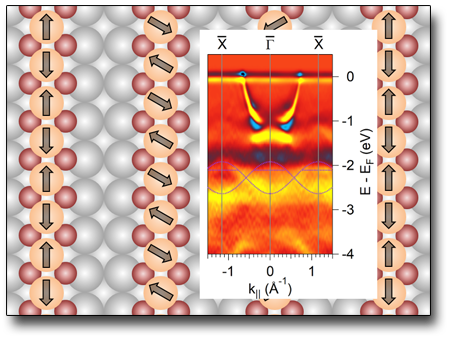
Manganese oxide chains grown on Ir(100) display a non-collinear spiral magnetic structure, which has been identified by spin-polarized scanning tunneling microscopy and theory. ARPES spectra show the effects on the Mn 3d states of the anti-ferromagnetic ordering along the chains, where the Mn atoms are adjacent.

See: M. Schmitt et al., Nat. Commun. 10, 2610 (2019)


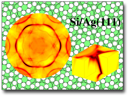
Electronic States of Silicene Allotropes on Ag(111)

Silicene is a honeycomb-like material similar to graphene and consisting of a single layer of silicon atoms. Some silicene allotropes can be synthesized on Ag(111). The ARPES analysis shows that these allotropic forms present σ-derived bands, while the π-derived bands expected in the proximity of the Fermi level are fully de-localized in Ag the substrate. This hybridization does not allow the formation of Dirac cones, at variance with the case of free-standing silicene.

See: P.M. Sheverdyaeva et al., ACS Nano 11, 975 (2017).


X-ray magnetic circular dichroism (XMCD)

X-Ray Magnetic Dichroism  (XMCD) is a differential absorption spectroscopy that exploits the interaction of x-ray circular polarized radiation with magnetic samples. It combines the information of the local sensitivity of core level excitation with the magnetic one. This spectroscopy provides the spin and the orbital magnetic moments of the atoms by means of the application of sum-rules.
In a single particle scenario, a two-step model provides an insight into the physical meaning:
1) electrons excited from core levels by circularly polarized light display an average spin momentum depending on the helicity,
2)  spin-polarized electrons probes in a different way the spin-resolved density of states above the Fermi level of the magnetized sample. The difference between the absorption taken at different helicity is related to the difference of the spin up and down branches of the density of empty states projected on the absorbing atoms and hence to the spin momentum of the absorbing atom.
XMCD spectroscopy efficiently probes the magnetic properties in the transition L2,3 metal edges (p -> d transition) and the rare-earths M4,5 edges (d -> f transition) because of the favorable optical selection rules which select the magnetic band as final state. 

CiPo@Elettra

X-ray magnetic circular dichroism

X-Ray Magnetic Dichroism  (XMCD) is a differential absorption spectroscopy that exploits the interaction of x-ray circular polarized radiation with magnetic samples. It combines the information of the local sensitivity of core level excitation with the magnetic one. This spectroscopy provides the spin and the orbital magnetic moments of the atoms by means of the application of sum-rules.
In a single particle scenario, a two-step model provides an insight into the physical meaning:
1) electrons excited from core levels by circularly polarized light display an average spin momentum depending on the helicity,
2)  spin-polarized electrons probes in a different way the spin-resolved density of states above the Fermi level of the magnetized sample. The difference between the absorption taken at different helicity is related to the difference of the spin up and down branches of the density of empty states projected on the absorbing atoms and hence to the spin momentum of the absorbing atom.
XMCD spectroscopy efficiently probes the magnetic properties in the transition L2,3 metal edges (p -> d transition) and the rare-earths M4,5 edges (d -> f transition) because of the favorable optical selection rules which select the magnetic band as final state.

CiPo@Elettra

X-ray absorption spectroscopy (XAS)

X-Ray Absorbtion Spectroscopy (XAS) is the extension of optical spectroscopy in the X-ray range. The X-ray energy range corresponds with the excitation of the core levels; due to the localized character of the core electron wave function the spectroscopic information is projected on the absorbing atom. Moreover, considering a single particle picture, since the core level has a sharp definition in energy and angular momentum, the XAS is proportional to the empty density of states projected on the absorbing atom and on the angular momentum l=±1, where l is the angular momentum of the core level. When this picture is no longer valid, i.e. effect of electron correlation are present, the interpretation of the spectra is closer to atomic-like multiplets.
The main advantage of XAS with respect to optical absorption is the chemical sensitivity via core level excitation. Soft X-ray Absorption is particularly suitable for C 1s, L2,3 of transition metals and M4,5 of rare-earths.
XAS display a great flexibility of experimental setups: it can be performed detecting transmitted photons, total and partial electron yield, fluorescence. For soft-X-rays the electron de-excitation channel is more efficient than the fluorescence mode.
Moreover, the direction of the linear polarization of the X-ray radiation with respect to the sample can give rise to change in the spectra by selection rules related to the dipole matrix element. This dependence is sensitive to anisotropy of charge and spin.

XAS@GasPhase XAS@VUV XAS@CiPo

Photon Induced Fluorescence Spectroscopy (PIFS)

PIFS is a photon in - photon out technique, exploiting characteristic "secondary" (or fluorescent) photons emitted from a material illuminated by X-rays.
This technique is available at the GasPhase beamline in two different variants that exploit bright, coherent, energy-tunable and monochromatic light form the Elettra synchrotron radiation source.
- X-ray Emission Spectroscopy (XES)
Thanks to its atomic sensitivity XES is a powerful analytical tool. It provides both site and element specificity, and it is extremely useful in investigating the electronic structure of molecules.The position of emission lines reflects small changes in occupied energy levels, whereas their intensity as a function of incident photon energy probes unoccupied molecular orbitals.  
- Vis-UV Fluorescence Spectroscopy
Analysis of vis-UV fluorescence allows to identify molecular and atomic photofragments resulting from relaxation dynamics of inner shell primary excitations. It allows thus to disentangle different relaxation mechanisms and obtain valuable informations on the nature of inner schell excitations.

PIFS

We use cookies essential for the functioning of the site. You can decide for yourself whether or not to allow cookies. Please note that if you refuse them, you may not be able to use all of the site's features.