UV-Vis Jasco2

Cell culture incubator, centrifuge, laminar flow hood and UV-VIS spectrophotometer

Marco Girasole - marco.girasole@ism.cnr.it

Giovanni Longo  - giovanni.longo@ism.cnr.it


BioTech@ISM

 
The support facility comprehend one cell culture incubator BINDER CB150 with two gas lines connected: nitrogen and carbon dioxide. One centrifuge Thermo MicroCl-17, not refrigerated, for the centrifugation of 1,5ml and 2ml test tubes in order to separate not only cells or bacteria but also organelles and proteins. The laminar flow hood allow the operator to safety work under sterile condition avoiding contamination from and to the ambient. It is equipped with activated carbon filters, one internal electric plug and a UV lamp to achieve a rapid sterilization. The spectrophotometer UV-VIS Jasco V-630 allow to rapidly measure solutions in standard cuvettes, it works in absorbance or transmission of light, in single or double (reference) beams configuration. It is also possible to work at fixed wavelength to follow in real-time enzymatic reactions.
A 4°C refrigerator and a -20°C freezer allow the storage of samples and reagents. A tabletop autoclave allows to sterilize materials for samples handlings. An high sensitivity analytical balance with automatic calibration allows to preparae buffers and solutions and two optical microscopes (one standard and one inverted) both equipped with objectives up to 50x allow for a rapid screening of the cultured cells and samples.
 

TECHNICAL SPECIFICATIONS

  • Cell culture incubator BINDER CB150 with temperature control (sensitivity 0,1 °C) and gas concentration control (sensitivity 0,1%)
  • 2 gas lines connected: CO2 e N2
  • Centrifuge with adjustable timer and rotation speed up to 3000 rpm
  • Laminar flow hood
  • Spectrophotometer Jasco V-630 UV/vis/NIR with double beam and single monochromator, wavelength range from 190 to 1100 nm.
  • Single beam working modality available as well as fixed wavelength time-course

AVAILABLE TECHNIQUES

  • UV-Vis spectroscopy
  • Centrifugation/Ultracentrifugation
 

SAMPLE

  • These equipment allow to maintain cells in culture under sterile conditions and to treat them in a safe way for the operator. Moreover it is possible to directly prepare solutions of interest and to rapidly characterize them with the spectrophotometer.


USE FOR

  • Measure of absorption, transmittance, reflectivity of samples in solution in the UV-Vis range

  • Measure of concentration of samples in solutioe

  • Measure of enzymatic activity

  • Measure of the progress of chemical reactions

  • Purify samples in solution by separating components of different molecular weights (proteins, cells, nanoparticles, surfactants etc.).


Case Studies

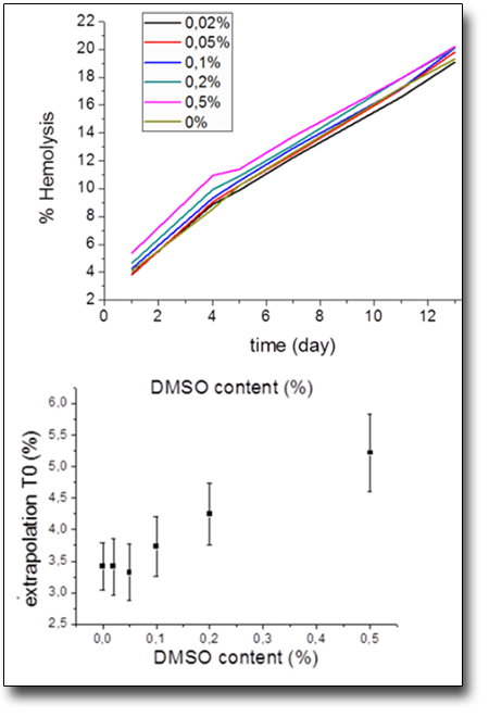
Toxic effect of DMSO on red blood cells

The dimethyl sulfoxide is well known in the literature to be a good deliverer of substances through the cellular plasma membrane, but, in high concentrations the drug intercalates the membrane massively and can create holes in the membrane itself. This issue is really important in red blood cells due to the lack of synthesis of enzymes that can remove the excess of dimethyl sulfoxide. In this sense we have followed complete ageing processes at different concentration of the DMSO in order to evaluate the most suitable working conditions. This evaluation has been carried out deriving the lysis percentage of the cells from the 350-700nm spectra of the supernatant of a sample’s aliquot at different days of ageing.


Evaluation of acetylcholinesterase (AChE) activity during in-vitro ageing of human red blood cells (RBC

Our preliminary data show that there isn’t a direct correlation between AChE activity and RBC ageing, in our experimental setup of accelerated in-vitro ageing in starvation condition. However our data show a sharp reduction of the enzyme activity after a process of “rejuvenation” of the cells in which in the incubation solution are added the necessary components to reactivate the metabolic cycle that synthetize ATP. This enzyme behavior is completely new and it calls for metabolic regulations and molecular mechanisms that we are actually studying.


X-ray photoelectron spectroscopy (XPS)

X-ray photoelectron spectroscopy (XPS) studies the chemical properties of solids through the photoelectric effect. In a XPS experiment a solid is irradiated with light of few tens to several hundred eV. The core-level electrons emitted from the sample carry element-specific information. By analyzing the energy spectrum of these electrons it is possible to detect the presence of atomic species, their chemical state and local bonding, their relative abundance (stoichiometry) and spatial distribution. The light sources used in XPS experiments range from X-ray tubes to synchrotrons that are optimized for different parts of the electromagnetic spectrum.

XPS@SAMOS XPS@Nanospectroscopy XPS/UPS@ONSET

UV-Vis spectroscopy

UV-VIS spectroscopy is a powerful analytical technique to determine the optical properties (transmittance, reflectance and absorbance) of liquids and solids.
It can be applied to characterize semiconductor materials, coatings, glass and many other research and manufacturing materials.  UV-Vis is often called a general technique because most molecules will absorb in the UV-Vis wavelength range. The UV extends from 100–400 nm and the visible spectrum from 400–700 nm.
When a photon hits a molecule and is absorbed, the molecule is promoted into a more excited energetic state. UV-visible light has enough energy to promote electrons to a higher electronic state, from the highest occupied molecular orbital (HOMO) to the lowest unoccupied molecular orbital (LUMO). The energy of the photon must exactly match the band gap for the photon to be absorbed. Thus, molecules with different chemical structures have different energy band gaps and different absorption spectra. The appearance of broad bands or shoulders on the UV-Vis structure is due to the numerous vibrational and rotational states of a molecule, which lead to separate energy band gaps of slightly different energies.

UV-Vis PerkinElmer Lambda950@RM UV-Vis Jasco1 UV-Vis Jasco2

Total X-ray fluorescence spectroscopy (TXRF)

Total X-rays Fluorescence (TXRF) is an ultra-sensitive surface elemental analysis technique. TXRF differs from other XRF techniques by its geometry: an incident beam of X-rays impinges upon a flat sample at angles below the critical angle of total external reflection, generally less than 1°. This grazing angle geometry allows the system to produce a relatively high signal-to-noise ratio and increase the surface sensitivity. The reflection of most of the excitation beam photons from the surface are collected on a detector. The software data analysis permits to identify the species on surface. It is possible to perform measurements on bulk, nanostructured and thin film samples.

Oxford-Si-Pin

Time-of-flight mass spectrometry (TOF MS)

Mass spectrometry (MS) is an analytical technique that measures the mass-to-charge (m/z) ratio of ions.
The experiment is performed by measuring the flight time of an ion from the ionization region to the detector and converting this quantity into m/z. The results are typically presented in the mass spectrum reporting the abundance of each ion versus the m/z ratio.

TOF